

 |  |
 | |
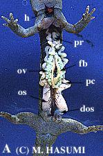
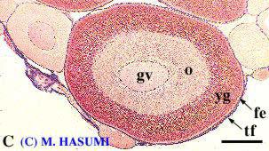
(A) August: remaining ovarian eggs colored mint green, perhaps to provide camouflage in green algae-laden ponds. At this time, vitellogenesis (i.e., accumulation of yolk granules) begins (see Fig. C), after which melanin deposition is initiated in the surface layer of the eggs in September.
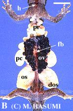
(B) October: the entire eggs are dark brown.
(C) Vitellogenic follicle from the August ovary (azan stain); fe = follicular epithelium, gv = germinal vesicle (nucleus), o = ooplasm, tf = theca folliculus, yg = yolk granules. Scale = 200 micrometer.
McDowell, W. T., and N. Shinozaki. 2015. A synopsis and larval description of Hynobius kimurae Dunn 1923 (Caudata: Hynobiidae). Bulletin of the Chicago Herpetological Society 50: 13-18.