| |
| |
|
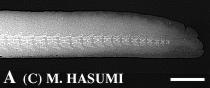
(A) Normal tail with normal and complete tail vertebrae. Tail tips or small parts of caudal fins are sometimes lost.
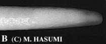
(B) Regenerated tail with partial or no tail vertebrae (see Fig. D).
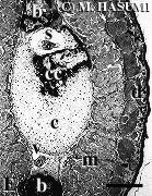
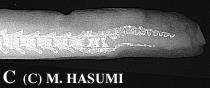
(C) Regenerated tail with incomplete or fragmentary tail vertebrae (see Fig. E).
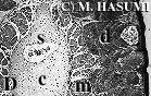
(D) Cross section of the regenerated tail having no vertebrae. Cartilage is observed to surround the spinal cord.
(E) Cross section of the regenerated tail with fragmentary vertebrae. Chondral cartilage or fragmentary bone tissue is discernible.
These regenerated tails are not distinguishable externally from those normal tails. The relative frequency of individuals with either externally broken tails or regenerated tails was much greater where oviposition sites were more rapidly flowing streams. This suggests that breeding adults of H. lichenatus, one of lentic-breeding hynobiids, do not so much adapt to lotic habitats.