| |
|
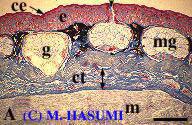
(A) Winter-dormant males during February (initial control). Note cornified epidermis, mucous glands filled with mucus, and a dense stratum (between arrows) of connective tissue under the skin. A very small amount of secreted mucus is recognized at the opening (arrow head) of the mucous glands. This is the condition of adult salamanders during the terrestrial phase (the surface of the skin is not sticky with mucus).
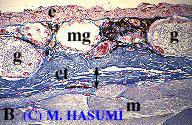
(B) Prolactin-injected males kept in a terrestrial habitat. Although the stratum of connective tissue under the skin (i.e., hypodermic tissue) is dense (between arrows), the mucous glands are completely empty. This result does not support the hypothesis that prolactin induces a remarkable enlargement of hypodermic tissue, causing animals to immigrate from land to water.
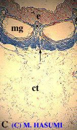
(C) Typical aquatic-phase males (for comparison). The hypodermic tissue is remarkably loose (under an arrow, 2-3 mm thick), and the mucous glands are empty.
Mucus in mucous glands is dyed dark blue with pH 2.5 alcian blue, suggesting it contains "acid mucopolysaccharides."