|
|
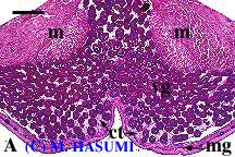
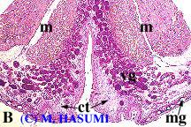
One type of a branched tubular gland, called "ventral gland," is found in the cloacal region.
(A) The ventral glands of males during spermiation, shortly after entering a pond, collected early in the March breeding season. Colloidal secretions, showing a strong PAS-positive reaction, are discernible in the lumina of many ventral glands. Mucous glands, distributed sporadically in the skin, are dyed blue with alcian blue.
(B) The ventral glands of males having a typical aquatic-phase morph, collected at the peak of the March breeding season. There are some secretory globules in the glandular lumina, epithelial cells keeping weakly PAS-positive color are mixed with unreactive ones, and the remarkable swelling of connective tissue is recognized under the skin. Note empty mucous glands.