
 |  |
 |  |
 |  |
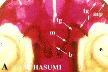
(A) Specimen with a deep bend toward the inside at the extreme border of a series of median prevomerine teeth (externally assuming U-shape), appeared in Tagami-machi, Niigata Prefecture, Japan. Such a type occurs generally in the specimens collected from the middle of the Niigata district.
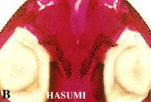
(B) Specimen with a small number of lateral prevomerine teeth, appeared in Takko-machi, Aomori Prefecture, Japan. This specimen has the greatest number of prevomerine teeth in this population.
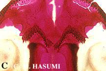
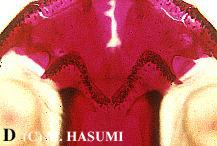
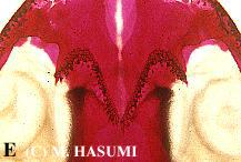
(C-E) Intrapopulation variation in the shape of a series of prevomerine teeth, appeared in Ohmagari-shi, Akita Prefecture, Japan. Especially, the specimen "E" has no bend toward the inside at the extreme border of a series of median prevomerine teeth (externally assuming V-shape). Clinal interpopulation variation is found in the relative frequency of such a type.
(F) Specimen with a series of median prevomerine teeth in which the right and left meet with each other, appeared in Minakami-machi, Gunma Prefecture, Japan.
One of old-fashioned taxonomic characters, U- or V-shape of prevomerine teeth, is no longer applicable to the classification of Hynobius species.
Visualization of the cranium stained with alizarinsulfonate (purple-red) through the transparent muscle of the specimen (alizarin stain).
*I employ here the term "prevomerine" according to Peters (1964): the prevomer has very often been used for the bone called the vomer in the palate of reptiles and amphibians because there is some question of its homology with the vomer of mammals, and vomero-palatine teeth are the same as vomerine teeth.
Peters, J. A. 1964. Dictionary of Herpetology. Hafner Publishing, New York, New York, U.S.A.